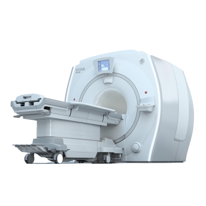
Cardiac Imaging
Hendrick Radiology Services offers noninvasive, comprehensive cardiac imaging to determine a wide range of cardiac needs. Available procedures include cardiac MRI, nuclear testing, ultrasound and computed tomography (CT). We provide imaging with the best low-contrast resolution at the lowest patient radiation dose.
-
James I.. Duff, MD
PathologyView Profile
-
John H.. Bliznak, MD
RadiologyView Profile
-
Michael J.. Maiers, MD
RadiologyView Profile
-
Steven J.. Nitke, MD
RadiologyView Profile
-
James A.. Rittimann, MD
Interventional RadiologyView Profile
-
Ashish C.. Patel, MD
RadiologyView Profile
-
Karen D.. Thomas, MD
PathologyView Profile
-
Ghassan A.. Tranesh, MD
PathologyView Profile
-
Jon T.. Anderson, MD
RadiologyView Profile
-
Ly T.. Ma, MD
PathologyView Profile


/
-
 Hendrick Imaging Center offers new ... Featured, Diagnostic Imaging & Radiology
Hendrick Health is offering the new preregistration process Virtual Intake Management℠(VIM) that gives patients the ability to complete registration ...
Continue Reading -
Cardiac MRI now available in ... Featured, Diagnostic Imaging & Radiology
Hendrick Health now offers cardiac MRI for advanced imaging applications of the heart and heart function. With the recent addition of software, ...
Continue Reading -
Stroke patient credits quick ... Featured, Diagnostic Imaging & Radiology
Phillip Brown cherishes more than ever the ability to hold his grandchildren. After experiencing a stroke, he feared he would be so debilitated that ...
Continue Reading -
Hendrick launches healthcare ... Diagnostic Imaging & Radiology
Hendrick Health continues to invest in the medical workforce of tomorrow with the recent launch of Healthcare Academy in partnership with Cisco ...
Continue Reading -
Billing/Business Services and ... Diagnostic Imaging & Radiology
Today, Hendrick Health relocated its customer services departments of Business Services/Billing and Medical Records/Release of Information to the ...
Continue Reading -
Hendrick Health announces ... Diagnostic Imaging & Radiology
Hendrick Health is pleased to announce the availability of DaTscan™ (Ioflupane I 123 Injection), the first FDA-approved imaging agent to help ...
Continue Reading -
Early detection for breast cancer ... Diagnostic Imaging & Radiology
Breast cancer is one of the most common cancers among American women, with one in eight women developing the disease in her lifetime. The American ...
Continue Reading -
Hendrick Health adds second linear ... Diagnostic Imaging & Radiology
As part of Hendrick Health’s commitment to innovative cancer care for the region, a second linear accelerator is now in operation at Hendrick Cancer ...
Continue Reading -
Hendrick Health brings advanced ... Diagnostic Imaging & Radiology
Hendrick Health is now offering new scanning technology that combines the advancements in magnetic resonance (MR) with the sophisticated engineering ...
Continue Reading -
Parking lot resurfacing project to ... Diagnostic Imaging & Radiology
Hendrick Medical Center, located at 1900 Pine St., will begin resurfacing patient parking lots on its campus on June 12. The project will affect, but ...
Continue Reading
-for-VINThrive-SQUARE.jpg)